Tuesday, December 30, 2008

HAEMOLYMPH

Haemolymph is the "blood" of insects. It is the watery fluid that fills the haemocoel. Haemolymph contains ions, molecules and cells. Often clear and colourless in most insects, some insects haemolymph may contain various pigments, making it appear yellow, blue, green, and in some rare cases of immature aquatic and endoparasitic flies, red due to the presence of haemoglobin.
All chemical exchanges between insect tissues are mediated through the haemolymph. The main difference between insect haemolymph and vertebrate blood is that haemolymph rarely contains respiratory pigments, and has a very low oxygen-transportation capacity. Respiration takes place in the tracheal system, which is mediated by the internal pressure of the haemolymph (the trachea opens and closes, creating a vacuum, as the haemolymph pressure changes).
Haemolymph is important to insect ventilation, thermoregulation, and molting (in breaking the old cuticle and expanding the new cuticle).
Haemolymph is a reserve of water for the insect. The soft-bodied insect larvae can be 20-40% haemolymph by weight, and the adult form is usually a bit less than 20% haemolymph (desiccation is always a problem for adult insects). The main constituent of haemolymph is plasma. Insect plasma is characterized by high concentrations of amino acids and organic phosphates.
Haemolymph not only provides nutrient transfer in the body, it provides protection for the insect. Haemolymph provides protection and defence from physical injury, disease organisms, parasites or other foreign objects entering the body, and sometimes from predation. In some insects, the haemolymph contains distasteful chemicals, which make the insect taste bad to predators (see aposematism). While an insect does have an immune system, please note that it is not as highly specialized as is the complex immunoglobin-based vertebrate immune system.
Blood Vessels


Blood vessels form a tubular network that allows blood to travels from the heart to the tissues and back to the heart again. Blood that leaves the heart passes into arteries. Large arteries branch into progressively smaller arteries that function to deliver blood to various regions of the body. Small arteries branch into even smaller vessels called arterioles, which function to regulate the flow of blood into different tissues. Arterioles branch into capillaries, the smallest of all blood vessels. Capillaries are the sites of nutrient and waste exchange between the blood and body cells. Capillaries are microscopic vessels that join the arterial system with the venous system. Blood coming out of the capillaries passes into vessels of increasing diameter as it flows back toward the heart. Capillaries join to form venules, which then merge to form small veins. Small veins unite to form large veins that eventually deliver blood back to the heart.

Arteries
Arteries serve as (1) efficient conduits for the movement of blood and (2) pressure reservoirs that keep blood moving during diastole. Arteries have a large internal diameter and thus offer little resistance to the flow of blood. Arteries also contain an elastic layer in their walls. Elastin is a protein fiber that has elastic qualities. During systole, large arteries distend with blood as their elastic walls stretch. During diastole, the walls rebound, thus pushing blood along. In this way the arteries act as a pressure reservoir that maintains a constant flow of blood through the capillaries despite pressure fluctuation during the cardiac cycle. Arteries also have a smooth muscular layer that functions to regulate the flow of blood through the artery. Contraction of the smooth muscle decreases the internal diameter of the vessel in a process called vasoconstriction. Relaxation of the smooth muscle increases the intermnal diameter in a process called vasodilation.

Arterioles
Arterioles serve as (1) the major determinant of blood pressure and (2) as the major determinant of blood flow to the individual organs Arterioles have a much smaller diameter than arteries and thus provide significant resistance to the flow of blood. This resistance creates pressure in the circulatory system. Pressure is required to provide adequate flow of blood to all parts of the body. Blood flow to individual organs can be regulated by controlling the diameter of the arterioles. Vasodilation of an arteriole lowers resistance and results in an increase in flow through that particular arteriole. Vasoconstriction of an arteriole increases resistance and results in decreased flow through that particular arteriole.

Capillaries

Capillaries are the smallest and most numerous of blood vessels. Capillaries function as the site of exchange of nutrients and wastes between blood and tissues. The anatomy of capillaries is well suited to the task of efficient exchange. Capillary walls are composed of a single layer of epithelial cells surrounded by a basement layer of connective tissue. The thin nature of the walls facilitates efficient diffusion of oxygen and carbon dioxide. Most capillaries also have pores between cells that allow for bulk transport of fluid and dissolved substances from the blood into the tissues and visa versa.
Although capillaries are extremely numerous (40 billion in the body), collectively they hold only about 5% of the total blood volume at any one time. This is because most capillaries are closed most of the time. Precapillary sphincters, which are bands of smooth muscle that wrap around arterioles, control the amount of blood flowing in a particular capillary bed. Contraction of the sphincter shuts off blood flow to a capillary bed, while relaxation of the sphincter allows blood to flow.

Veins

Veins are larger and more compliant (stretchable) than arteries, thus they can hold more blood. In fact, the veins act somewhat like a blood reservoir, containing 60% of the total blood volume at rest. As physical activity increases, the veins undergo vasoconstriction, driving more blood back to the heart and increasing circulation. Also, the return of venous blood to the heart is aided by one-way valves that insure unidirectional flow of blood


HEART ATTACK

A heart attack occurs when blood flow to a section of heart muscle becomes blocked. If the flow of blood isn’t restored quickly, the section of heart muscle becomes damaged from lack of oxygen and begins to die.

Heart attacks occur most often as a result of a condition called coronary artery disease (CAD). In CAD, a fatty material called plaque (plak) builds up over many years on the inside walls of the coronary arteries (the arteries that supply blood and oxygen to your heart). Eventually, an area of plaque can rupture, causing a blood clot to form on the surface of the plaque. If the clot becomes large enough, it can mostly or completely block the flow of oxygen-rich blood to the part of the heart muscle fed by the artery.


Heart With Muscle Damage and a Blocked Artery


Figure A is an overview of a heart and coronary artery showing damage (dead heart muscle) caused by a heart attack. Figure B is a cross-section of the coronary artery with plaque buildup and a blood clot.
During a heart attack, if the blockage in the coronary artery isn’t treated quickly, the heart muscle will begin to die and be replaced by scar tissue. This heart damage may not be obvious, or it may cause severe or long-lasting problems.
Severe problems linked to heart attack can include heart failure and life-threatening arrhythmias (irregular heartbeats). Heart failure is a condition in which the heart can’t pump enough blood throughout the body. Ventricular fibrillation is a serious arrhythmia that can cause death if not treated quickly.

Get Help Quickly

Acting fast at the first sign of heart attack symptoms can save your life and limit damage to your heart. Treatment is most effective when started within 1 hour of the beginning of symptoms.
The most common heart attack signs and symptoms are:
Chest discomfort or pain—uncomfortable pressure, squeezing, fullness, or pain in the center of the chest that can be mild or strong. This discomfort or pain lasts more than a few minutes or goes away and comes back.
Upper body discomfort in one or both arms, the back, neck, jaw, or stomach.
Shortness of breath may occur with or before chest discomfort.
Other signs include nausea (feeling sick to your stomach), vomiting, lightheadedness or fainting, or breaking out in a cold sweat
EMBOLISM


Definition
An embolism is an obstruction in a blood vessel due to a blood clot or other foreign matter that gets stuck while traveling through the bloodstream. The plural of embolism is emboli.


Description
Emboli have moved from the place where they were formed through the bloodstream to another part of the body, where they obstruct an artery and block the flow of blood. The emboli are usually formed from blood clots but are occasionally comprised of air, fat, or tumor tissue. Embolic events can be multiple and small, or single and massive. They can be life-threatening and require immediate emergency medical care. There are three general categories of emboli: arterial, gas, and pulmonary. Pulmonary emboli are the most common.

Arterial embolism
In arterial emboli, blood flow is blocked at the junction of major arteries, most often at the groin, knee, or thigh. Arterial emboli are generally a complication of heart disease. An arterial embolism in the brain (cerebral embolism) causes stroke, which can be fatal. An estimated 5-14% of all strokes are caused by cerebral emboli. Arterial emboli to the extremities can lead to tissue death and amputation of the affected limb if not treated effectively within hours. Intestines and kidneys can also suffer damage from emboli.

Gas embolism
Gas emboli result from the compression of respiratory gases into the blood and other tissues due to rapid changes in environmental pressure, for example, while flying or scuba diving. As external pressure decreases, gases (like nitrogen) that are dissolved in the blood and other tissues become small bubbles that can block blood flow and cause organ damage.

Pulmonary embolism


In a pulmonary embolism, a common illness, blood flow is blocked at a pulmonary artery. When emboli block the main pulmonary artery, and in cases where there are no initial symptoms, a pulmonary embolism can quickly become fatal. According to the American Heart Association, an estimated 600,000 Americans develop pulmonary emboli annually and 60,000 die from it.
A pulmonary embolism is difficult to diagnose. Less than 10% of patients who die from a pulmonary embolism were diagnosed with the condition. More than 90% of cases of pulmonary emboli are complications of deep vein thrombosis, blood clots in the deep vein of the leg or pelvis.

Causes and symptoms
Arterial emboli are usually a complication of heart disease where blood clots form in the heart's chambers. Gas emboli are caused by rapid changes in environmental pressure that could happen when flying or scuba diving. A pulmonary embolism is caused by blood clots that travel through the blood stream to the lungs and block a pulmonary artery. More than 90% of the cases of pulmonary embolism are a complication of deep vein thrombosis, which typically occurs in patients who have had orthopedic surgery and patients with cancer or other chronic illnesses like congestive heart failure.

Risk factors for arterial and pulmonary emboli include: prolonged bed rest, surgery, childbirth, heart attack, stroke, congestive heart failure, cancer, obesity, a broken hip or leg, oral contraceptives, sickle cell anemia, chest trauma, certain congenital heart defects, and old age. Risk factors for gas emboli include: scuba diving, amateur plane flight, exercise, injury, obesity, dehydration, excessive alcohol, colds, and medications such as narcotics and antihistamines.

Symptoms of an arterial embolism include:
severe pain in the area of the embolism
pale, bluish cool skin
numbness
tingling
muscular weakness or paralysis

Common symptoms of a pulmonary embolism include:
labored breathing, sometimes accompanied by chest pain
a rapid pulse
a cough that may produce sputum
a low-grade fever
fluid build-up in the lungs

Less common symptoms include:
coughing up blood
pain caused by movement or breathing
leg swelling
bluish skin
fainting
swollen neck veins

Monday, November 24, 2008

COMPOSITION OF BLOOD


Components of Blood - average adult has about 5 liters (about 5 qts):

1 - Formed elements:
Red blood cells (or erythrocytes)
White blood cells (or leucocytes)
Platelets (or thrombocytes)

2 - Plasma = water + dissolved solutes

Red blood cell, platelet, and white blood cell


Red Blood Cells (or erythrocytes):
1 - biconcave discs
2 - lack a nucleus & cannot reproduce (average lifespan = about 120 days)
3 - transport hemoglobin (each RBC has about 280 million hemoglobin molecules)
4 - Typical concentration is 4-6 million per cubic mm (or hematocrit [packed cell volume] of about 42% for females & 45% for males)
5 - contain carbonic anhydrase (critical for transport of carbon dioxide)

Hemoglobin


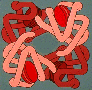
Composed of globin (made up of 4 highly folded polypeptide chains) + 4 heme groups (with iron)
each molecule can carry 4 molecules of oxygen
called oxyhemoglobin when carrying oxygen & called reduced hemoglobin when not carrying oxygen
can also combine with carbon dioxide & helps transport carbon dioxide from the tissues to the lungs


White blood cells (or leucocytes or leukocytes):
Have nuclei & do not contain hemoglobin
Typical concentration is 5,000 - 9,000 per cubic millimeter
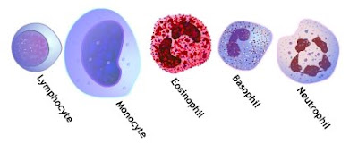
Types of WBCs:
granular white blood cells include:
neutrophils (50 - 70% of WBCs)
eosinophils (1 - 4%)
basophils (less than 1%)
agranular (or non-granular) white blood cells include:
lymphocytes (25 - 40%)
monocytes (2 - 8%)

Granular white blood cells contains numerous granules in the cytoplasm, & their nuclei are lobed.
Agranular white blood cells have few or no granules in the cytoplasm & have a large spherical nucleus.

Granular white blood cells are produced in the bone marrow, while agranular white blood cells are produced in lymph tissue, e.g., Lymph nodes (specialized dilations of lymphatic tissue which are supported within by a meshwork of connective tissue called reticulin fibers and are populated by dense aggregates of lymphocytes and macrophages).

The primary functions of the various white blood cells are:

Neutrophils - phagocytosis (bacteria & cellular debris); very important in inflammation


Eosinophils - help initiate and sustain inflammation and can activate T-cells (directly by serving as antigen-presenting cells and indirectly by secreting a variety of cytokines. Eosinophils can also kill bacteria by quickly releasing mitochondrial DNA and proteins (described below).
Eosinophils respond to diverse stimuli, including tissue injury, infections, allografts, allergens, and tumors. Eosinophils can also release a variety of cytokines, chemokines, lipid mediators, and neuromodulators. Eosinophils directly communicate with T cells and mast cells. Eosinophils activate T cells by serving as antigen-presenting cells.

Basophils - along with mast cells, play a role in inflammation and allergic responses
Release of histamine (that contributes to the 'symptoms' of allergies) by mast cells requires the production of antibodies (IgE) by B-cells andthat process is regulated, in part, by cytokines produced by basophils (Bischoff 2007).

Monocytes - phagocytosis (typically as macrophages in tissues of the liver, spleen, lungs, & lymph nodes) & also important antigen-presenting cells
Once distributed through the blood stream, monocytes enter other tissues of the body such as the liver (Kupffer cells), lungs (alveolar macrophages), skin (Langerhans cells), and central nervous system (microglia) (Gordon 2003).

Lymphocytes - immune response (including production of antibodies)

Eosinophils (in green with red nucleus) catapult their mitochondrial DNA out of the cell, forming tangled traps (red) that ensnare foreign bacteria.


Platelets (or thrombocytes)
1 - formed in the bone marrow from cells called megakaryocytes
2 - have no nucleus, but can secrete a variety of substances & can also contract (because they contain actin & myosin)
3 - normal concentration in the blood is about 250,000 per cubic millimeter
4 - remain functional for about 7 - 10 days (after which they are removed from the blood by macrophages in the spleen & liver)
5- play an important role in hemostasis (preventing blood loss)

Plasma:
1 - Water - serves as transport medium; carries heat
2 - Proteins
Albumins
60-80% of plasma proteins
most important in maintenance of osmotic balance
produced by liver

Globulins
alpha & beta
some are important for transport of materials through the blood (e.g., thyroid hormone &
iron)
some are clotting factors
produced by liver
gamma globulins are immunoglobulins (antibodies) produced by lymphocytes

Fibrinogen
important in clotting
produced by liver

3 - Inorganic constituents (1% of plasma) - e.g., sodium, chloride, potassium, & calcium
4 - Nutrients - glucose, amino acids, lipids & vitamins
5 - Waste products - e.g., nitrogenous wastes like urea
6 - Dissolved gases - oxygen & carbon dioxide
7 - Hormones
HOW DOES THE HEART BEAT


The atria and ventricles work together, alternately contracting and relaxing to pump blood through your heart. The electrical system of your heart is the power source that makes this possible.
Your heartbeat is triggered by electrical impulses that travel down a special pathway through your heart.
SA node (sinoatrial node) - known as the heart's natural pacemaker The impulse starts in a small bundle of specialized cells located in the right atrium, called the SA node. The electrical activity spreads through the walls of the atria and causes them to contract.
AV node (atrioventricular node) The AV node is a cluster of cells in the center of the heart between the atria and ventricles, and acts like a gate that slows the electrical signal before it enters the ventricles. This delay gives the atria time to contract before the ventricles do.
His-Purkinje Network This pathway of fibers sends the impulse to the muscular walls of the ventricles and causes them to contract.
At rest, a normal heart beats around 50 to 99 times a minute. Exercise, emotions, fever and some medications can cause your heart to beat faster, sometimes to well over 100 beats per minute.

Tuesday, November 18, 2008

LYMPHATIC SYSTEM


The lymphatic system consists of the tissues and organs that produce, store, and carry lymphocytes (a type of white blood cell), which fight infections and other diseases. The lymphatic system includes the bone marrow, spleen, thymus, lymph nodes, and lymphatic vessels (a network of thin tubes that carry lymph and white blood cells, or leukocytes). Lymphatic vessels branch, like blood vessels, into all the tissues of the body. Functions of the lymphatic system
Formation of the interstitial fluid As blood passes through the capillaries some of the water-soluble material materials and white cells escape through the capillary walls. This liquid fills the space between the tissue cells and constitues the interstitial fluid, or tissue fluid. Part of the institial fluid is absorbed back into the capillaries. The rest passes into the lymph vessels to become lymph.

The lymphatic system has three main functions.

First, it returns excess interstitial fluid (also called tissue fluid) to the blood. Of the fluid that leaves the capillaries, about 90 percent is returned. The 10 percent that doesn't return becomes part of the tissue fluid that surrounds the tissue cells. Small protein molecules may "leak" through the capillary wall and increase the osmotic pressure of the interstitial fluid. This further inhibits the return of fluid into the capillaries, and fluid tends to accumulate in the tissue spaces. If this continues, blood volume and blood pressure decrease significantly and the volume of tissue fluid increases, which results in edema (swelling). Lymph capillaries pick up the excess interstitial fluid and proteins and return them to the venous blood. After the fluid enters the lymph capillaries, it is called lymph.

The second function of the lymphatic system is the absorption of fats and fat-soluble vitamins from the digestive system and the subsequent transport of these substances to the venous circulation. The mucosa that lines the small intestine is covered with fingerlike projections called villi. There are blood capillaries and special lymph capillaries, called lacteals (see below), in the center of each villus. The blood capillaries absorb most nutrients, but the fats and fat-soluble vitamins are absorbed by the lacteals. The lymph in the lacteals has a milky appearance due to its high fat content and is called chyle.

The third and probably most well known function of the lymphatic system is defense against invading microorganisms and disease. In other words, the lymph system is part of the immune system. Lymph nodes and other lymphatic organs filter the lymph to remove microorganisms and other foreign particles. Lymphatic organs contain lymphocytes that destroy invading organisms.

The usual function of the lymphatics is to carry lymph from the tissue back to the bloodstream, but the great neywork of lymphatic vessels which serves the intestines also transports nutrient substances from the food. During digestion the small molecules into which food is broken down fron dtheir way into the millions of tiny villi which line the interior of the intestinal wall. Some of these food molecules are carried away in the bloodstream, but others, particularly fats, enter the tiny lymphatic vessles, called lacteals, one of which lies in the center of each villus. The mixture of lymph and nutrients, known as chyle, is carried by the lacteals to lymph vessels in the intestinal wall. It is then collected into the larger vessels which course through the mesentery and is carried to the cisterna chyli. Cisterna chyli The cisterna chyli is an irregularly shaped chamber about 6 cm (2½ inches) long and about 2.5 cm (1 inch) wide which lies on the rear wall of the abdominal cavity. The intestinal lymph trunk, which carries the lymph from the intestines, and the left and right lumbar trunks, which carry lymph from the lower limbs and the pelvis, lead into it. At its upper end it narrows to form the lower end of the thoracic duct.

Thoracic duct
The thoracic duct carries the mixture of lymph and chyle from the cisterna chyli upwards through the thorax to the left subclavian vein in the root of the neck. It is the largest lymph vessel in the body, being about 41 cm (16 inches) long and 0.6 (0.25 inch) wide. Changes in pressure in the body, associated with breathing, cause the lymph to move up through the thoracic duct, which is provided with several valves to prevent the fluid from flowing downwards. Just before the thoracic duct enters the subclavian vein it is joined by the left jugular and subclavian trunks carrying lymph from the left side of the head and neck and the left arm.

Right lymphatic duct
The lymph from the right side of the head and neck is collected into the right jugular trunk, while that from the right arm flows into the right subclavian trunk. These vessels unite to form the right lymphatic duct which carries the lymph to the right subclavian vein.

Lymph nodes
A lymph node and section of a lymph vessel
The lymph vessels which drain the lymph from the tissues do not run directly to the large lymph vessels, but are interrupted in their courses by small organs known as lymph nodes. These lymph nodes are the "glands" which can often be felt under the skin. Their function is to filter the lymph, removing from it any dangerous microbes which may have found their way through the skin or the intestinal wall and into the interstitial spaces. In most cases several afferent lymph vessels carry lymph into the outer part of each lymph node. The lymph passes through the mode and is carried away from it in one or more efferent lymph vessels, and so on to the next node. And so the lymph makes its way by steps to the thoracic duct or the right lymphatic duct. The lymph vessels which carry the lymph from node to node are extremely thin-walled and delicate. Externally they have a beaded appearance. This caused by the presence within of minute one-way valves.

Disorders of the lymphatic system
In some conditions, such as after radical mastectomy, the lymphatic vessels to a limb become obstructed, causing lymph to accumulate and the limb to become hard and swollen, a condition known as lymphoedema. Cancer commonly spreads via the lymphatic system. A primary tumor invades the lymphatics and fragments of tumor (metastases) break off and travel to the local group of lymph nodes, where the metastases continue to grow and produce a secondary tumor.

CARDIOVASCULAR SYSTEM


Also known as the cardiovascular system, the system that, in humans and other higher animals, delivers oxygen and nutrients throughout the body by a complex network of vessels – arteries, arterioles, capillaries, veins, and venules. Arteries, arterioles, and the microscopic capillaries carry blood away from the heart to all parts of the body and allow exchange of nutrients and wastes through capillary walls from blood to the tissues and organs. Veins carry deoxygenated blood back to the lungs for reoxygenation. If all the vessels of this network in an adult human body were laid end-to-end, they would extend for about 60,000 miles (more than 96,500 kilometers) – far enough to circle the Earth more than twice. As in the adult, survival of the developing embryo depends on the circulation of blood to maintain homeostasis and a favorable cellular environment. In response to this need, the circulatory system makes its appearance early in development and reaches a functional state long before any other major organ system. Incredible as it seems, the primitive heart begins to beat regularly early in the fourth week following fertilization. The vital role of the circulatory system in maintaining homeostasis depends on the continuous and controlled movement of blood through the thousands of miles of capillaries that permeate every tissue and reach every cell in the body. It is in the microscopic capillaries that blood performs its ultimate transport function. Nutrients and other essential materials pass from capillary blood into fluids surrounding the cells as waste products are removed. Numerous control mechanisms help to regulate and integrate the diverse functions and component parts of the cardiovascular system in order to supply blood to specific body areas according to need. These mechanisms ensure a constant internal environment surrounding each body cell regardless of differing demands for nutrients or production of waste products.

Circulatory pathways
The blood vessels of the body are functionally divided into two distinctive circuits: pulmonary circuit and systemic circuit. The pump for the pulmonary circuit, which circulates blood through the lungs, is the right ventricle. The left ventricle is the pump for the systemic circuit, which provides the blood supply for the tissue cells of the body.

Pulmonary circulation transports oxygen-poor blood from the right ventricle to the lungs where blood picks up a new blood supply. Then it returns the oxygen-rich blood to the left atrium. Systemic Circuit The systemic circulation provides the functional blood supply to all body tissue. It carries oxygen and nutrients to the cells and picks up carbon dioxide and waste products.

Systemic circulation carries oxygenated blood from the left ventricle, through the arteries, to the capillaries in the tissues of the body. From the tissue capillaries, the deoxygenated blood returns through a system of veins to the right atrium of the heart.

The coronary arteries are the only vessels that branch from the ascending aorta. The brachiocephalic, left common carotid, and left subclavian arteries branch from the aortic arch. Blood supply for the brain is provided by the internal carotid and vertebral arteries. The subclavian arteries provide the blood supply for the upper extremity. The celiac, superior mesenteric, suprarenal, renal, gonadal, and inferior mesenteric arteries branch from the abdominal aorta to supply the abdominal viscera. Lumbar arteries provide blood for the muscles and spinal cord. Branches of the external iliac artery provide the blood supply for the lower extremity. The internal iliac artery supplies the pelvic viscera.

Major systemic arteries
All systemic arteries are branches, either directly or indirectly, from the aorta. The aorta ascends from the left ventricle, curves posteriorly and to the left, then descends through the thorax and abdomen. This geography divides the aorta into three portions: ascending aorta, arotic arch, and descending aorta. The descending aorta is further subdivided into the thoracic arota and abdominal aorta. Major systemic veins After blood delivers oxygen to the tissues and picks up carbon dioxide, it returns to the heart through a system of veins. The capillaries, where the gaseous exchange occurs, merge into venules and these converge to form larger and larger veins until the blood reaches either the superior vena cava or inferior vena cava, which drain into the right atrium.

Fetal circulation
Most circulatory pathways in a fetus are like those in the adult but there are some notable differences because the lungs, the gastrointestinal tract, and the kidneys are not functioning before birth. The fetus obtains its oxygen and nutrients from the mother and also depends on maternal circulation to carry away the carbon dioxide and waste products. The umbilical cord contains two umbilical arteries to carry fetal blood to the placenta and one umbilical vein to carry oxygen-and-nutrient-rich blood from the placenta to the fetus. The ductus venosus allows blood to bypass the immature liver in fetal circulation. The foramen ovale and ductus arteriosus are modifications that permit blood to bypass the lungs in fetal circulation.

LEARN ABOUT CARDIOVASCULAR SYSTEM

Click here to learn how the cardiovascular system works